Diagnostics
Leading the way in technology for vision loss and blindness.
VMR Institute has been on the forefront of developing and implementing novel diagnostic technologies not found elsewhere, affording patients the latest in imaging and vision function testing.
[divider scroll_text=””]
Standard Clinical Testing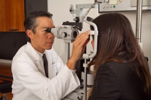
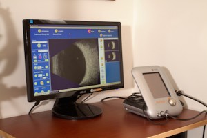
The VMR Institute is equipped with the latest imaging technologies that are all computerized and networked. This means that you don’t need to go elsewhere for testing, since it can all be done in the office, usually on the same day as your visit. Because all tests are computerized, the results are available immediately. In almost all cases, you will leave the office with an answer.
[toggle_box]
[toggle_item title=”Standard Testing” active=”true”]
Standard testing includes state-of-the-art digital imaging for retinal and choroidal angiography, digital ultrasonography, and combined optical coherence tomography – scanning laser ophthalmoscopy.
[/toggle_item]
[toggle_item title=”Digital Fundus Photography “]
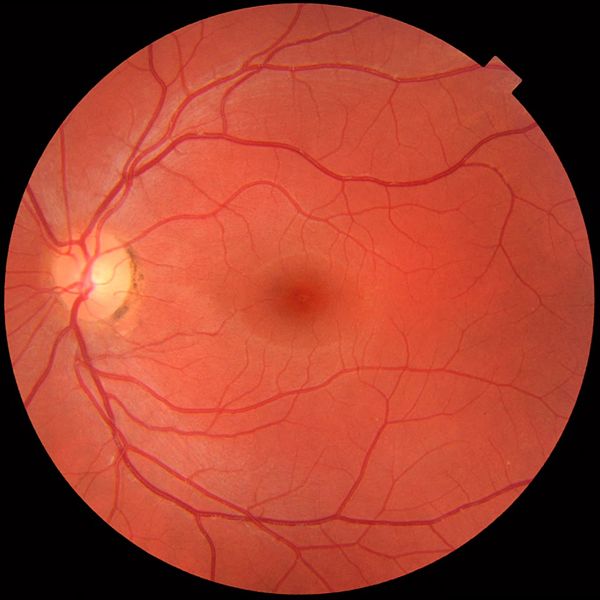
Digital Fundus Photography allows for photographing of the interior parts of the eye including the retina, macula and optic disc in order to better detect, and monitor, conditions such as diabetic retinopathy, macular degeneration, and disorders of the macula and retinal vasculature.
[/toggle_item]
[toggle_item title=”Digital Fluorescein Angiography“]
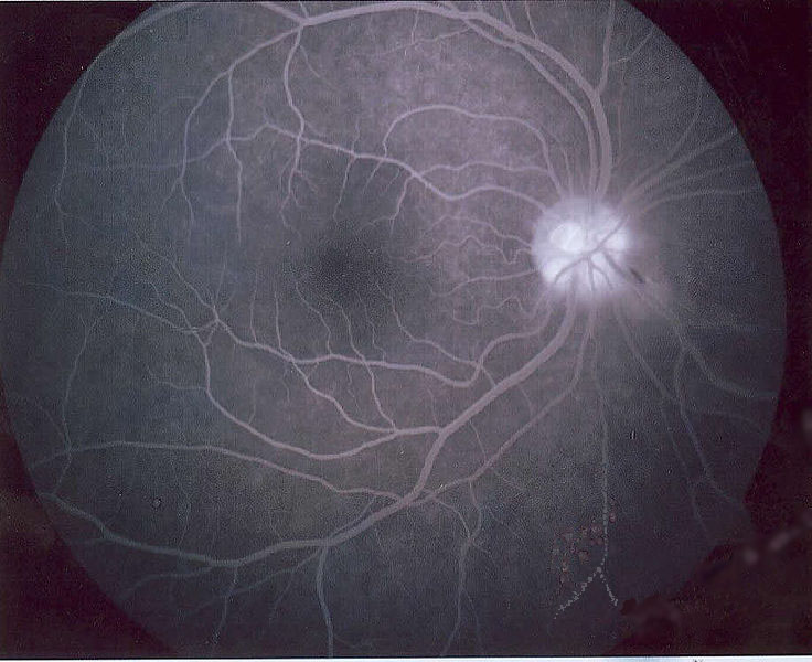
Digital Fluorescein Angiography is a technique used to examine the eye by which a fluorescent dye is injected into the eye and then, using a specialized camera, enables the doctor to observe and thus make accurate diagnoses and determine if laser surgery or drug injections can treat diabetic retinopathy, macular degeneration, and other retinal disorders.
[/toggle_item]
[toggle_item title=”Digital Ophthalmic Ultrasonography“]
Digital Ophthalic Ultrasonography is used to evaluate floaters, retinal detachments, tumors, intraocular foreign bodies, and trauma.
[/toggle_item]
[toggle_item title=”Combined Optical Coherence Tomography (OCT) – Scanning Laser Ophthalmoscopy (SLO) “]
The VMR Institute was among the first centers in the world to use this new imaging technology that provides much greater detail than ever before.
[/toggle_item]
[/toggle_box]
[divider scroll_text=””]
Novel Diagnostic Technologies
For years, the VMR Institute has been involved in developing new ways to diagnose problems in the Vitreous, Macula, & Retina. These programs have been in collaboration with scientists at prestigious institutions across the country.
NASA Collaborative Research
In collaboration with Dr. Rafat Ansari of the NASA Glenn Research Center in Cleveland, Dr. Sebag is helping to develop new diagnostic instrumentation to assist in detecting diseases of the vitreous, macula, and retina early in their course. The earlier a disease is diagnosed, the more effective will be the treatment. Since the eye is the window to the body, this advanced new technology will not only enable non-invasive diagnostics of eye diseases, but also diseases of the brain and body. NASA hopes to use this new diagnostic technology on astronauts in space, but at the VMR Institute the same technology will be available to patients participating in research studies.
[toggle_box]
[toggle_item title=”View related VMR Publications to learn more:” active=”true”]
Ansari RR, Rovati L, Sebag J: Celestial and terrestrial tele-ophthalmology: a health monitoring helmet for astronauts/cosmonauts and general public use. Ophthalmic Technologies XI (Manns, Soderberg, Ho; eds). Proc SPIE 245:177-85, 2001
Ansari RR, Dunker S, Suh K, Kitaya N, Sebag J: Quantitative molecular characterization of bovine vitreous and lens with non-invasive dynamic light scattering. Exp Eye Res 73:859-866, 2001
[/toggle_item]
[/toggle_box]
Doheny Eye Institute Collaborative Research on Floaters
![]()
Recent studies in collaboration with Alfredo Sadun, MD, PhD of the Doheny Eye Institute have led to a deeper understanding of why floaters disturb vision. Using advanced methods of measuring vision, we have identified that patients suffering from floaters are unhappy because contrast sensitivity is cut in half. Fortunately, this can be cured by minimally-invasive vitrectomy and contrast sensitivity returns to normal.
[toggle_box]
[toggle_item title=”View related VMR Publications to learn more:” active=”true”]
[/toggle_item]
[/toggle_box]
CalTech /Jet Propulsion Laboratory Collaborative Research
Dr. Sebag and Dr. Sadun have collaborated with Dr. Terri Lawton and Dr. Wolfgang Fink to develop prototype equipment to measure vision at the VMR Institute in Huntington Beach. Many of these tests are not available elsewhere. The results of this research have provided new insights into how various diseases disturb vision. Some examples are:
[toggle_box]
[toggle_item title=”View related VMR Publications to learn more:” active=”true”]
Lawton TA, Sebag J, Sadun AA, Castleman KR: Image enhancement improves reading performance in age-related macular degeneration patients. Vision Res 38:153-162, 1998
Jivrajka R V, Kim J K, Fink W, Sadun AA, Sebag J: Quantitative analysis of central visual field defects in macular edema using three-dimensional computer-automated threshold Amsler grid testing. Graefes Arch Clin Exp Ophthalmol 247:165-70, 2009
Tozer K, Fink W, Sadun AA, Sebag J: Prospective three-dimensional analysis of structure and function in macular hole treated by pharmacologic vitreolysis.
Retinal Cases & Brief Reports 7: 57-61, 2013.
[/toggle_item]
[/toggle_box]